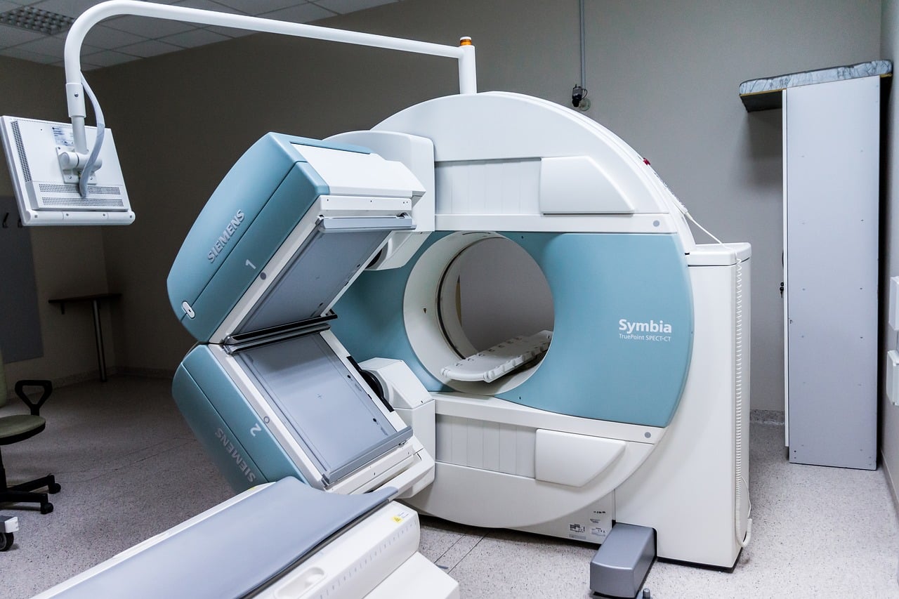
Scientists are looking into ways to make MRI scans as safe as possible, and artificial intelligence (AI) may hold the key. Researchers now suggest it may be possible to use AI for future MRI scans to reduce the amount of contrast dye needed for the test.
What is gadolinium?
Gadolinium, a contrast dye often used to enhance the images doctors receive from MRIs, is a heavy metal. Previous studies have found that some gadolinium remains in the bodies of patients after they undergo an MRI with contrast.
“There is concrete evidence that gadolinium deposits in the brain and body,” lead author and Stanford University researcher Enhao Gong, Ph.D. said in a statement. “While the implications of this are unclear, mitigating potential patient risks while maximizing the clinical value of the MRI exams is imperative.”
Although the impact of gadolinium on humans is still unknown, radiologists are working toward a way to reduce the amount needed for the scan while enhancing patients’ safety. Now researchers say using AI for future MRI scans seems like the best way to reduce the amount of gadolinium while still capturing images with the fine details physicians need to see from an MRI with contrast.
Artificial intelligence is accurate
There is a wealth of potential applications for AI in medicine and science, which is why Dr. Gong and his team studied deep learning and neural networks to find a solution to this problem. They used machine learning models called convolutional neural networks, which allow the computer to recognize images created through MRI scanning while observing distinctions in scanning the data. Scientists believe AI could see the data in ways a “human observer might not be capable of discerning.”
After the training, the machine learning algorithm could make approximations and recognize distinctions between full-dose scans and zero-dose and low-dose MRIs. The results suggested the image quality didn’t differ drastically between the low-dose, algorithm-enhanced MRIs and the MRIs done with a full dose of contrast.
No drastic change in quality
Thanks to this study, neurologists could soon consider switching to AI for future MRI scans in place of MRIs with contrast.
“Low-dose gadolinium images yield significant untapped clinically useful information that is accessible now by using deep learning and AI,” Dr. Gong said.
Nevertheless, this approach requires more studying and evaluation of the algorithm using more MRI scanners and other contrast agents, which the team plans to do in the future.
“We’re not trying to replace existing imaging technology,” Dr. Gong said. “We’re trying to improve it and generate more value from the existing information while looking out for the safety of our patients.”